


Last updated on June 24th, 2020
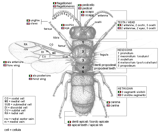
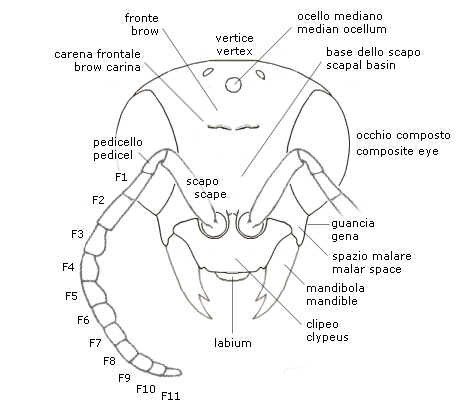
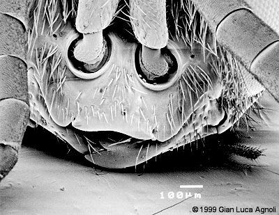
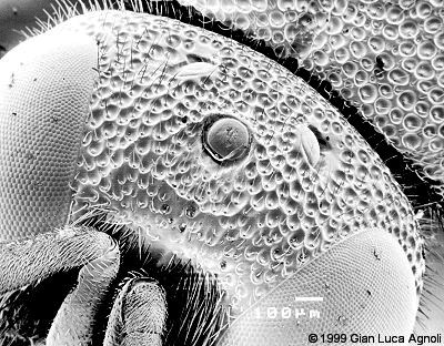
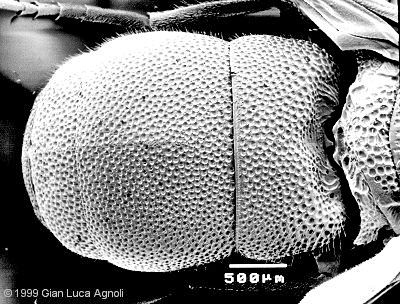
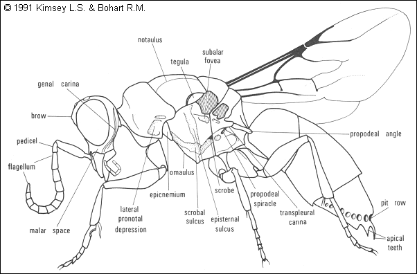
The HEAD is hypognathous, so the mouth apparatus is turned downwards. Two composed eyes and three simple ocelli are present, on the vertex. The space between the base of the eyes and the articulation of the jaws is the malar space, of taxonomic importance. The antennae are inserted little above the jaws, in correspondence of the upper part of the clypeus: here are the scape, the pedicel and 11 flagellomeres (F-1, F-2, etc.), generally cylindrical. The relative length of the first 3 flagellomeres is of diagnostic importance. The clypeus is generally short and wide, except for the Stilbum Genus. The jaws are simple and are armed with 0-3 apical teeth. The central part of the face is occupied by a depression (scapal basin) able to receive the antennas in a folded position. The upper part of the face is the forehead, often characterized by a crossing carina projecting behind to the area of the ocelli (vertex).
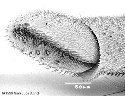
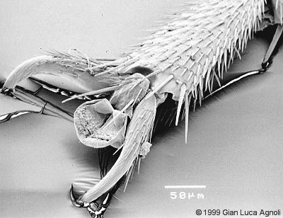
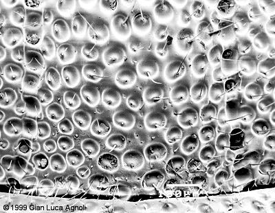
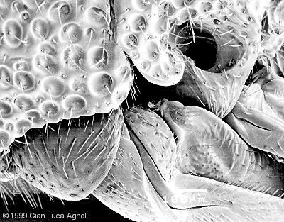
The THORAX is subdivided into 5 articulate segments: pronoto, mesonotum (scutum), scutellum, metanotum (postscutellum) and propodeum. The surficial sculpture can be absent (smooth teguments) or intense (punctuation, drills, carinas). To the pronotum, comprising the neck, articulates the first pair of legs. The mesonotum (scutum) is characterized by 3 regions (two lateral and one central = mesoscutum or medium mesonotum separated by notauli and laterally by the 2 tegulae that cover the wing articulation. The scutellum is generally shorter than the scutum. The metanotum can be characterized by a projection (Elampus, Parnopes), or can be fused with the propodeum; it is often characterized by the presence of lateral metathoracic teeth. The propodeum, precisely the first gastral segment, is generally vertical and lacks an upper flat face; it’s also characterized by two lateral teeth. The legs are generally adapted for an excavating activity. The segments that compose the legs are the hip (coxa), the femur, the tibia and the tarsus. The terminal part of the tarsus is armed with dentate nails.
In modern hymenopterological nomenclature the terms mesosoma and metasoma are used instead of thorax and abdomen, respectively. The reason is that the first abdominal segment (propodeum) is joint with the morphological thorax and the famous “wasp-waist” articulates two abdominal segments and not the thorax to the abdomen.
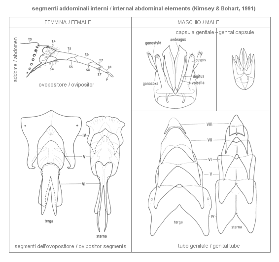
 The ABDOMEN of Chrysidids is significantly modified when compared to the abdomen of the other Hymenoptera, and is generally reduced to only 3 visible segments and other internal segments.The Amiseginae, Cleptinae and Loboscelidiinae have 5 external abdominal segments in males and 4 in females, the Parnopinae have 4 in males and 3 in females, the Allocoeliini 2 tergites (dorsal segments) and 3 sternites (ventral segments) in both sexes. In the other tribes males and females have 3 abdominal segments.The external segments are generally very sclerotized and concave in the ventral side, except for Cleptinae, Amiseginae and Loboscelidiinae, where the sternites are convex.A useful diagnostic structure is found in the anal edge of the last tergite, characterized by teeth, carinas, projections and spots.Above the anal edge a characteristic pit row is often present. The internal abdominal segments form a subcylindric telescopic tube, introflected during the rest, representing the female’s ovipositor and the male’s genital tube. The sting sensu strictu is reduced and is no more working. In males, at the end of the tube a genital capsula is present, standing on the eighth sternite, both structures of diagnostic importance.
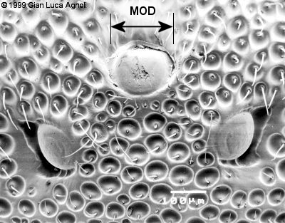
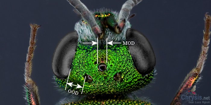
When measuring morphological characters under a microscope, the following measures are used:
MOD, the Diameter of the Median Ocellus. This quantity is used as the unit for all the other measures taken on a given specimen, which become relative measures (eg. 2.0 MOD, 4.5 MOD, etc.) instead of absolute measures (eg. 3 millimeters). If measures are expressed in MOD units, and since the Diameter of the Median Ocellus is proportional to the size of the specimen, you can compare the same measures obtained on different specimens in order to draw average values and other statistics.
OOD (Ocellar-Ocular Distance), the minimum distance between lateral ocellus and compound eye.
PD (Puncture Diameter), the diameter of the punctuation on a given area, eg. on the pronotum, on the T-2, etc.
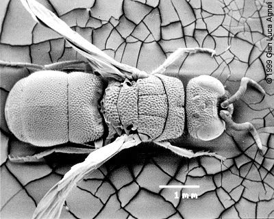
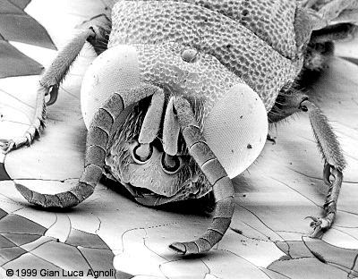
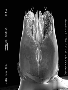
All the micrographs were taken with a JEOL JSM-5400 Scanning Electron Microscope, after a vacuum ionization of the dried specimen (Chrysis scutellaris) with gold (JVG-N1 Gauge and JEE-4B Evaporator).
Copyright, Authorship, and Ownership statements
All text and images of this page are copyright ©️ Chrysis.net unless otherwise stated - please see individual cases for authorship and copyright details. The specimens pictured are from the authors' or other collaborators' personal collections and from the collections of various museums. Unless otherwise specified, the whole content of this web site is for personal, non-commercial, scientific, and educational purposes given proper accreditation to the page from which they were derived are provided, and under Chrysis.net Terms and Conditions.
For citation purposes
Agnoli G.L. & Rosa P. (2026) Morphology of Chrysididae, in: Chrysis.net website. Interim version 20 July 2026, URL: https://www.chrysis.net/chrysididae/morphology-of-chrysididae/.